Causes: In 1992, Paul Ridker at Brigham and Women's Hospital began asking a question that the cardiology establishment found puzzling. Many people who had heart attacks had normal cholesterol levels. What if cholesterol wasn't the whole story?
What if the real culprit was something more fundamental - something that cholesterol was merely a symptom of?
He started measuring C-reactive protein (CRP), a molecule synthesized by the liver in response to inflammatory signaling, in blood samples that had been collected years before patients' cardiac events.
What he found was striking: people with elevated CRP faced dramatically higher heart attack risk - even when their cholesterol was normal. Ridker wasn't finding a new risk factor. He was finding evidence for a new model of how heart disease works.
The JUPITER trial, published in the New England Journal of Medicine in 2008, tested his hypothesis. He randomized 17,802 people with low LDL but elevated CRP to either a statin (rosuvastatin) or placebo. The statin reduced both LDL and CRP.
The result: 44% reduction in cardiovascular events. More striking still, the CANTOS trial in 2017 tested an antibody (canakinumab) that blocked the inflammatory cytokine IL-1beta - with no effect on cholesterol levels at all. It reduced cardiovascular events.
Inflammation wasn't just associated with heart disease. It was causing it.
The same case has been made, with varying degrees of evidence, for a remarkable list of conditions: type 2 diabetes, Alzheimer's disease, depression, certain cancers, metabolic syndrome, chronic kidney disease, and autoimmune conditions.
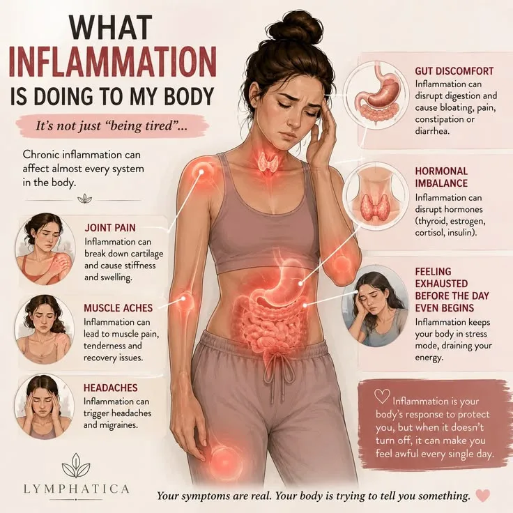
Chronic low-grade inflammation - not the dramatic, purposeful inflammation of a healing wound, but a persistent, smoldering activation of immune signaling that damages tissue over years - has become one of the central concepts in the biology of modern chronic disease.
"Inflammation is a fire that we set to protect ourselves. But we're living in a world that keeps the fire going all the time." - Andrew Miller, Emory University
Key Definitions
Acute inflammation - The immune system's rapid, targeted response to tissue damage or infection. Characterized by redness, swelling, heat, pain, and loss of function at the site. Pro-inflammatory: mediated by prostaglandins, leukotrienes, histamine, and cytokines (IL-1, IL-6, TNF-alpha).
Self-limiting: resolves when the triggering stimulus is cleared.
Chronic low-grade inflammation - Persistent, systemic, mild elevation of pro-inflammatory markers without an acute infectious or injury cause. Typically detected by elevated serum CRP, IL-6, TNF-alpha, or fibrinogen. Subclinical - does not produce the obvious signs of acute inflammation - but damages tissue over decades.
Cytokines - Small signaling proteins secreted by immune cells that regulate immune responses. Pro-inflammatory cytokines (TNF-alpha, IL-1, IL-6) amplify immune activation; anti-inflammatory cytokines (IL-10, TGF-beta) resolve it.
Chronically elevated pro-inflammatory cytokines at low levels are the biochemical signature of chronic inflammation.
C-reactive protein (CRP) - A plasma protein synthesized by the liver in response to IL-6 signaling; used clinically as a marker of systemic inflammation.
High-sensitivity CRP (hs-CRP) distinguishes levels in the low chronic range (0.5-3 mg/L, associated with cardiovascular risk) from the high acute range (>10 mg/L, indicating active infection or injury).
NF-kB (Nuclear factor kappa B) - A master transcription factor that activates hundreds of pro-inflammatory genes. Activated by a wide range of stimuli including LPS (bacterial cell wall components), oxidized LDL, cytokines, ROS (reactive oxygen species), and psychosocial stress hormones.
A convergence point for many chronic inflammation drivers.
Inflammaging - Claudio Franceschi's term for the chronic, low-grade inflammatory state that develops with aging. Driven by accumulated senescent cells (which secrete pro-inflammatory SASP), mitochondrial dysfunction, cumulative gut dysbiosis, and decades of chronic inflammation drivers.
A primary mechanism of age-related disease and functional decline.
Intestinal permeability - The degree to which the gut barrier allows passage of molecules from the intestinal lumen into systemic circulation. Compromise of the gut barrier ("leaky gut") allows bacterial LPS to translocate into blood, driving systemic TLR4-mediated inflammatory activation.
Adipokines - Signaling molecules secreted by adipose (fat) tissue. Visceral fat is metabolically active, secreting pro-inflammatory adipokines (leptin, resistin, IL-6, TNF-alpha) at higher rates than subcutaneous fat. The pro-inflammatory character of visceral adipose tissue is a primary mechanism connecting obesity to chronic disease.
Advanced glycation end products (AGEs) - Molecules formed when proteins or fats are exposed to sugars (glycation) or high heat. AGEs accumulate in the body with elevated blood glucose and dietary intake; they activate the RAGE receptor, driving NF-kB-mediated inflammation.
Implicated in diabetic complications and the inflammatory effects of ultra-processed foods.
Omega-3/omega-6 balance - Omega-3 fatty acids (EPA, DHA from fatty fish; ALA from flaxseed) are metabolic precursors to anti-inflammatory eicosanoids. Omega-6 fatty acids (linoleic acid, primarily from seed oils) are precursors to pro-inflammatory eicosanoids.
Modern Western diets have dramatically shifted from the 4:1 or lower omega-6:omega-3 ratio estimated for evolutionary human diets to ratios of 15:1 or higher, with pro-inflammatory consequences.
The Mechanism: From Trigger to Tissue Damage
Chronic inflammation follows a common mechanistic pathway regardless of its specific trigger:
1. Triggering signals: PRR (pattern recognition receptor) activation, particularly TLR (toll-like receptor) activation by LPS or other pathogen-associated molecular patterns (PAMPs), or DAMP (damage-associated molecular pattern) activation by cellular stress signals. Both converge on NF-kB activation.
2. NF-kB activation: NF-kB translocates to the nucleus and activates transcription of pro-inflammatory cytokines (TNF-alpha, IL-1beta, IL-6), chemokines (attracting immune cells), adhesion molecules (recruiting circulating immune cells to the tissue), and cyclooxygenase-2 (COX-2, producing prostaglandins).
3. Macrophage polarization: Macrophages shift from M2 (anti-inflammatory, reparative) to M1 (pro-inflammatory) phenotype, producing more cytokines, reactive oxygen species, and proteolytic enzymes that damage tissue.
4. Tissue damage: Chronically activated immune cells produce reactive oxygen species (ROS) and nitrogen species, metalloproteinases, and inflammatory mediators that damage structural proteins, DNA, and cell membranes. This damage generates more DAMPs, feeding back into the activation loop.
5. Systemic effects: Liver production of acute phase proteins (CRP, fibrinogen, serum amyloid A) elevates; endothelial dysfunction occurs; insulin resistance develops; neural signaling via the vagus nerve and blood-brain barrier transmits inflammatory signals to the brain.
The Major Disease Connections
Cardiovascular Disease: The Inflammatory Plaque
The inflammatory model of atherosclerosis is now the dominant mechanistic framework in cardiology.
The process begins not with cholesterol accumulation but with endothelial activation: pro-inflammatory stimuli (oxidized LDL, elevated glucose, mechanical stress, elevated cytokines) activate the endothelium, which expresses adhesion molecules that recruit monocytes from the blood.
These monocytes differentiate into macrophages in the arterial intima, engulf oxidized LDL particles, and transform into foam cells - the defining cellular component of the fatty streak that begins plaque formation.
The foam cells and activated endothelium secrete cytokines that recruit more monocytes, smooth muscle cells, and T cells, establishing a self-amplifying inflammatory lesion.
Peter Libby's foundational work identified that vulnerable plaques - those most likely to rupture and cause myocardial infarction - are distinguished by active inflammation: thin fibrous caps, abundant macrophages, and metalloproteinase activity that degrades the structural proteins holding the plaque together.
Many large, calcified plaques never rupture; many smaller, inflamed plaques do.
Ridker's CANTOS trial - providing the first direct human evidence for anti-inflammatory therapy in cardiovascular prevention - used canakinumab to block IL-1beta. The trial found 15% reduction in cardiovascular events with no change in lipid levels.
Anti-inflammatory therapy works for cardiovascular disease through pathways entirely independent of cholesterol.
Type 2 Diabetes: Fat, Inflammation, and Insulin Resistance
The mechanistic connection between visceral obesity, chronic inflammation, and insulin resistance is among the most thoroughly characterized in metabolic biology.
Visceral adipocytes (fat cells in the abdominal depot) are metabolically distinct from subcutaneous fat. They are larger, more lipolytically active, and surrounded by macrophages that produce pro-inflammatory cytokines.
TNF-alpha, produced by both adipocytes and infiltrating macrophages, directly inhibits insulin receptor signaling - it phosphorylates the insulin receptor substrate (IRS-1) in a way that blocks the downstream signaling cascade that allows glucose uptake.
This mechanism, identified by Gokhan Hotamisligil in the 1990s, established that inflammation directly causes insulin resistance at the molecular level. The cycle is self-amplifying: visceral fat drives inflammation, which drives insulin resistance, which drives hyperglycemia, which produces AGEs and ROS, which drive more inflammation.
The implication is that anti-inflammatory strategies - exercise (which reduces visceral fat and has direct anti-inflammatory effects), dietary changes, and sleep improvement - can improve insulin sensitivity through inflammatory mechanisms, not just through caloric effects on body composition.
Alzheimer's Disease: Neuroinflammation as Driver
The recognition that neuroinflammation is a primary driver of Alzheimer's - not merely a reactive consequence - has fundamentally reshaped the field.
Genome-wide association studies (GWAS) for late-onset Alzheimer's disease have identified multiple susceptibility genes, and strikingly, many of the most significant ones are specifically expressed in microglia (the brain's resident immune cells): TREM2, CD33, CR1, CLU.
These are not neuronal genes - they are immune genes. The genetic architecture of Alzheimer's disease is substantially an immune disease architecture.
Microglia in Alzheimer's disease shift from neuroprotective (clearing amyloid and cellular debris) to pro-inflammatory phenotypes that secrete cytokines, produce ROS, and damage synapses and neurons.
TREM2 loss-of-function variants, which impair microglial phagocytic capacity, are one of the strongest single-gene risk factors for late-onset Alzheimer's after APOE4.
The peripheral inflammation connection: systemic pro-inflammatory cytokines communicate to the brain via the blood-brain barrier (cytokines can cross directly when the barrier is compromised) and via the vagus nerve.
Systemic infections, gut dysbiosis, and metabolic inflammation all appear to accelerate amyloid accumulation and cognitive decline - suggesting that treating peripheral inflammation may have neuroprotective effects.
Depression: Inflammation and the Sad Brain
Andrew Miller and Charles Raison's work on inflammation and depression provides one of the strongest arguments that a subset of depressive illness is fundamentally an inflammatory disease.
The natural experiment: interferon-alpha (IFN-alpha) treatment for hepatitis C and certain cancers produces clinical depression in 40-50% of patients within weeks of initiation. The dose, timing, and reversibility are consistent and reproducible.
Administration of IFN-alpha also activates the IDO (indoleamine 2,3-dioxygenase) enzyme, which diverts tryptophan metabolism from serotonin synthesis toward kynurenine production - directly reducing brain serotonin while generating quinolinic acid, a neurotoxic NMDA receptor agonist that damages hippocampal neurons.
The observational correlate: multiple meta-analyses find significantly elevated CRP, IL-6, and TNF-alpha in depressed patients compared to healthy controls. The elevation is not merely explained by shared risk factors like obesity or sedentary behavior - it appears independently.
The clinical implications: patients with treatment-resistant depression and elevated inflammatory markers have been studied with TNF-alpha inhibitors (infliximab) and celecoxib (a COX-2 inhibitor). In subsets of patients with elevated CRP, both show antidepressant effects.
This suggests that inflammatory-subtype depression may be a pharmacologically distinct condition better treated by anti-inflammatory approaches than by monoaminergic manipulation.
The Gut: The Inflammation Gateway
The gut microbiome's role in chronic inflammation has emerged as one of the most consequential findings of the past two decades. Three specific mechanisms connect gut biology to systemic inflammation:
Intestinal Permeability and LPS Translocation
The intestinal epithelium is a single-cell-layer barrier between the gut lumen - which contains trillions of bacteria and their products - and the systemic circulation. The barrier is maintained by tight junction proteins (occludin, claudins, zonulin) that seal the space between epithelial cells.
When the gut barrier is compromised - by dysbiosis, ultra-processed food consumption, alcohol, NSAID overuse, or chronic stress - bacterial LPS (lipopolysaccharide) from gram-negative bacteria translocates from the gut into the blood.
LPS activates TLR4 on macrophages and other immune cells with extraordinary potency, triggering robust NF-kB-mediated inflammatory responses.
Cani and colleagues at the Catholic University of Louvain documented this mechanism comprehensively: high-fat diet in mice produces gut dysbiosis, increased intestinal permeability, elevated plasma LPS (called metabolic endotoxemia), and systemic inflammation - with the sequence running from the gut outward.
Short-Chain Fatty Acids: The Anti-Inflammatory Microbiome Output
Butyrate, propionate, and acetate - short-chain fatty acids (SCFAs) produced by fermentation of dietary fiber by gut bacteria - have potent anti-inflammatory effects. Butyrate is the primary energy source for colonocytes (colon cells), maintaining intestinal barrier integrity.
It also inhibits NF-kB directly, reduces production of pro-inflammatory cytokines, and promotes regulatory T cell differentiation.
The dramatic decline in dietary fiber consumption in Western populations - estimated at 15-20 g/day compared to estimated evolutionary intake of 50-100+ g/day - has substantially reduced SCFA production and the microbial communities that produce them.
This fiber gap may be one of the most important contributors to the chronic inflammation driving modern chronic disease.
Microbiome Diversity and Immune Calibration
The gut microbiome educates the immune system during development and continues to calibrate its baseline tone throughout life. Reduced microbiome diversity - consistently found in populations eating Westernized diets - is associated with elevated inflammatory markers, increased intestinal permeability, and reduced SCFA production.
Eran Segal and Eran Elinav's work at the Weizmann Institute documented remarkable inter-individual variation in gut microbiome composition and glycemic responses to identical foods - establishing the microbiome as a major determinant of metabolic response that may explain why population-level dietary recommendations produce variable individual results.
What Reduces Chronic Inflammation
The evidence converges on several categories of intervention with meaningful anti-inflammatory evidence:
Exercise
Muscle contractions during exercise produce IL-6 from working muscle - but this IL-6 acts paradoxically as an anti-inflammatory myokine (rather than its pro-inflammatory role in adipose tissue): it stimulates anti-inflammatory IL-10 production and inhibits TNF-alpha.
Regular aerobic exercise consistently reduces resting CRP and IL-6 levels, reduces visceral fat (the primary adipose inflammation driver), and reduces macrophage infiltration into adipose tissue.
The anti-inflammatory effect of exercise is dose-dependent: even moderate amounts (30 minutes/day, most days) produce measurable CRP reduction in clinical trials.
Diet
The Lyon Diet Heart Study (1999) - secondary prevention after myocardial infarction - compared a Mediterranean-style diet to a prudent Western diet. Five-year cardiovascular mortality was reduced by 73% in the Mediterranean group. No pharmaceutical trial has produced equivalent effect sizes.
The anti-inflammatory components of the Mediterranean diet include:
- Olive oil: oleocanthal inhibits COX-1 and COX-2 (the aspirin mechanism); polyphenols inhibit NF-kB
- Omega-3 fatty acids (EPA, DHA from fatty fish): converted to anti-inflammatory resolvins, protectins, and maresins
- Dietary fiber: feeds SCFA-producing bacteria, improves gut barrier, reduces LPS translocation
- Polyphenols (vegetables, legumes, wine): multiple anti-inflammatory mechanisms via Nrf2 and NF-kB modulation
Ultra-processed food avoidance works through eliminating AGEs (from dry-heat processed foods), trans fats, high omega-6:omega-3 ratios, and refined carbohydrates that drive glucose-NF-kB activation.
Sleep
A single night of partial sleep deprivation (4 hours instead of 8) elevates TNF-alpha by 0.31-0.69 pg/mL and produces measurable NF-kB activation in circulating monocytes. Chronic short sleep (6 vs 8 hours) maintains elevated CRP and IL-6.
The mechanism involves elevated evening cortisol, SNS activation impairing sleep quality, and reduced nocturnal cellular repair processes.
Stress Management
Psychosocial stress drives inflammation through HPA axis (cortisol at high levels paradoxically upregulates inflammatory gene expression through GR-mediated resistance mechanisms) and SNS (norepinephrine activates adrenergic receptors on immune cells, upregulating NF-kB).
MBSR has documented reductions in CRP and inflammatory gene expression profiles in small trials. Social connection reduces the CTRA inflammatory gene expression profile.
| Intervention | Effect on Inflammation | Mechanism | Evidence Strength |
|---|---|---|---|
| Aerobic exercise | Reduces CRP, IL-6 | Myokine signaling, visceral fat reduction | Strong RCTs |
| Mediterranean diet | Reduces CRP, oxidized LDL | Polyphenols, omega-3s, fiber | Strong RCTs and cohorts |
| Sleep (7-9 hrs) | Reduces CRP, IL-6 | Reduced cortisol/SNS activation | Experimental studies |
| Not smoking | Reduces CRP dramatically | Removes primary ROS/oxidative stress | Strong observational |
| Visceral fat reduction | Reduces TNF-alpha, IL-6 | Reduces adipokine secretion | Mechanistically robust |
| Omega-3 supplementation | Modestly reduces CRP | EPA/DHA to resolvins | Modest RCT effects |
| Probiotic/fiber increase | Reduces LPS, improves barrier | Gut microbiome | Emerging evidence |
The Inflammaging Problem
As people age, chronic inflammation becomes self-amplifying in a way that captures all the above mechanisms.
Cellular senescence - the accumulation of damaged cells that stop dividing but remain metabolically active - produces the SASP (senescence-associated secretory phenotype): a cocktail of pro-inflammatory cytokines, metalloproteinases, and other mediators that damage surrounding tissue and recruit macrophages.
Senescent cell accumulation is one of the most important drivers of inflammaging.
Mitochondrial dysfunction with age produces reactive oxygen species (ROS) that activate NF-kB. Gut microbiome diversity declines with age, reducing SCFA production. Decades of cumulative environmental inflammation drivers (diet, stress, infections) compound.
The adaptive immune system becomes "exhausted," with reduced ability to mount specific responses while innate immune activation (source of cytokine-driven chronic inflammation) remains high.
Senolytics - drugs that selectively clear senescent cells - are the most promising anti-inflammaging intervention in development. Dasatinib + quercetin and navitoclax both reduce SASP markers and improve healthspan in animal models; human clinical trials are underway.
For related concepts, see how the human immune system works, why diets fail, how stress damages the body, what the science of longevity shows, and how the gut microbiome works.
Sources & Further Reading
- Ridker, P. M., et al. (2008). Rosuvastatin to Prevent Vascular Events in Men and Women with Elevated C-Reactive Protein. New England Journal of Medicine, 359(21), 2195-2207. DOI: 10.1056/NEJMoa0807646
- Ridker, P. M., et al. (2017). Antiinflammatory Therapy with Canakinumab for Atherosclerotic Disease. New England Journal of Medicine, 377(12), 1119-1131. DOI: 10.1056/NEJMoa1707914
- Libby, P. (2002). Inflammation in Atherosclerosis. Nature, 420(6917), 868-874. DOI: 10.1038/nature01323
- Hotamisligil, G. S., Shargill, N. S., & Spiegelman, B. M. (1993). Adipose Expression of Tumor Necrosis Factor-alpha: Direct Role in Obesity-Linked Insulin Resistance. Science, 259(5091), 87-91. DOI: 10.1126/science.7678183
- Miller, A. H., & Raison, C. L. (2016). The Role of Inflammation in Depression: From Evolutionary Imperative to Modern Treatment Target. Nature Reviews Immunology, 16(1), 22-34. DOI: 10.1038/nri.2015.5
- Cani, P. D., et al. (2007). Metabolic Endotoxemia Initiates Obesity and Insulin Resistance. Diabetes, 56(7), 1761-1772. DOI: 10.2337/db06-1491
- de Lorgeril, M., et al. (1999). Mediterranean Diet, Traditional Risk Factors, and the Rate of Cardiovascular Complications After Myocardial Infarction. Circulation, 99(6), 779-785. DOI: 10.1161/01.CIR.99.6.779
- Franceschi, C., & Campisi, J. (2014). Chronic Inflammation (Inflammaging) and Its Potential Contribution to Age-Associated Diseases. Journals of Gerontology, 69(Suppl 1), S4-S9. DOI: 10.1093/gerona/glu057
Frequently Asked Questions
What is the difference between acute and chronic inflammation?
Acute inflammation is the immune system’s rapid, targeted response to injury or infection: redness, swelling, heat, and pain at the site of damage, produced by a coordinated cascade of prostaglandins, cytokines (IL-1, IL-6, TNF-alpha), and immune cell recruitment. It is self-limiting, once the threat is resolved, anti-inflammatory signals terminate the response and repair begins. Chronic inflammation is different in kind, not just duration. It is typically low-grade, systemic rather than local, and persists without an acute infectious or injury cause. Chronic inflammation features mildly elevated pro-inflammatory cytokines (CRP, IL-6, TNF-alpha at levels insufficient to produce obvious symptoms but sufficient to damage tissue over years and decades). It does not resolve because the stimuli driving it, metabolic stress, gut dysbiosis, visceral fat, persistent infection, psychosocial stress, are chronic rather than acute. The distinction matters because acute inflammation is protective and necessary; chronic inflammation is the shared mechanism underlying most chronic diseases of aging.
What are the main drivers of chronic inflammation?
The major drivers of chronic low-grade inflammation cluster in several categories. Visceral (abdominal) fat is one of the most potent: adipocytes (fat cells), particularly in the visceral depot, secrete pro-inflammatory cytokines (adipokines including leptin, resistin, and IL-6) and activate macrophages that infiltrate adipose tissue and produce TNF-alpha. Gut dysbiosis and intestinal permeability allow bacterial lipopolysaccharide (LPS), a component of gram-negative bacterial cell walls, to translocate from the gut into systemic circulation, triggering chronic TLR4-mediated immune activation. Ultra-processed food consumption drives inflammation through multiple pathways: advanced glycation end products (AGEs), trans fats, refined carbohydrate-driven glucose spikes, and displacement of anti-inflammatory omega-3 fatty acids with pro-inflammatory omega-6 fatty acids. Psychosocial stress activates the SNS and HPA axis, producing cortisol and catecholamines that upregulate the NF-kB inflammatory transcription pathway. Sleep deprivation elevates pro-inflammatory cytokines. Sedentary behavior reduces anti-inflammatory mechanisms normally activated by muscle contraction. Environmental toxins, air pollution, and dysbiosis contribute further.
How does inflammation cause heart disease?
The inflammatory model of atherosclerosis, developed by Peter Libby, Paul Ridker, and others, replaced the older view of heart disease as simple cholesterol plaque accumulation with a much richer mechanistic picture. The process begins when oxidized LDL particles infiltrate the arterial wall and trigger an inflammatory response: macrophages are recruited, engulf the oxidized LDL to become ‘foam cells,’ and secrete pro-inflammatory cytokines that perpetuate the process. Smooth muscle cells migrate and proliferate, forming the fibrous cap over the growing plaque. The vulnerable plaques that cause myocardial infarctions are not necessarily the largest ones, they are the most inflamed ones, with thin caps and active macrophage infiltration that make them prone to rupture. CRP (C-reactive protein), synthesized by the liver in response to IL-6, became the key clinical marker for cardiovascular inflammation. Paul Ridker’s JUPITER trial (2008) found that statin treatment reduced cardiovascular events even in people with low LDL but elevated CRP, and more recently, his CANTOS trial (2017) found that canakinumab (an IL-1beta antibody with no lipid-lowering effects) reduced cardiovascular events, directly proving the inflammatory mechanism.
What is the connection between inflammation and depression?
The inflammatory theory of depression, developed most comprehensively by Andrew Miller and Charles Raison, proposes that chronic activation of the immune system produces depressive symptoms through direct effects on neurotransmitter metabolism and brain function. The evidence includes: (1) Administration of interferon-alpha (IFN-alpha) to treat hepatitis C causes depression in 40-50% of patients, a controlled human model of inflammation-induced depression; (2) People with depression have significantly elevated inflammatory markers (CRP, IL-6, TNF-alpha) compared to non-depressed controls; (3) Anti-inflammatory treatments (including NSAIDs, celecoxib, and TNF-alpha inhibitors used for rheumatological conditions) show antidepressant effects in patients with elevated inflammation; (4) The IDO (indoleamine 2,3-dioxygenase) enzyme, activated by inflammation, converts tryptophan to kynurenine rather than serotonin, directly reducing serotonin synthesis while generating neurotoxic quinolinic acid. The practical implication: the subset of depressed patients with elevated inflammatory markers may respond preferentially to anti-inflammatory treatments rather than monoaminergic antidepressants, a potential basis for depression subtypes that could guide treatment selection.
How does inflammation contribute to Alzheimer's disease?
Neuroinflammation has moved from being considered a consequence of Alzheimer’s disease to being recognized as an active driver of it. Microglia, the brain’s resident immune cells, normally clear amyloid-beta and other metabolic waste. In Alzheimer’s disease, chronically activated microglia become pro-inflammatory rather than clearing: they secrete cytokines (IL-1beta, TNF-alpha, IL-6) that damage neurons and impair synaptic function. Genome-wide association studies have identified multiple Alzheimer’s risk genes, including TREM2, CD33, CR1, and CLU, that are specifically expressed in immune cells and involved in neuroinflammatory regulation, establishing a direct genetic link. The discovery that systemic inflammation exacerbates brain amyloid accumulation (LPS injection increases brain amyloid in mouse models) suggests that peripheral inflammation communicates to the brain through the blood-brain barrier and vagus nerve, accelerating neurodegeneration. This has potential therapeutic implications: several anti-inflammatory strategies are in Alzheimer’s clinical trials.
What dietary changes most effectively reduce chronic inflammation?
The Mediterranean diet has the most evidence for anti-inflammatory effects: abundant olive oil (oleocanthal, an ibuprofen-like COX inhibitor), vegetables and legumes (polyphenols, fiber, and prebiotics), fatty fish (omega-3 fatty acids EPA and DHA), and moderate wine (resveratrol). The Lyon Diet Heart Study, a secondary prevention trial of Mediterranean diet after heart attack, found a 73% reduction in cardiac events, a magnitude larger than any pharmaceutical trial in cardiovascular prevention. Ultra-processed food avoidance is the most powerful single dietary change: Hall’s NIH RCT found that ultra-processed diet consumption increased multiple inflammatory markers, while unprocessed diet reduced them, even when macronutrients were matched. Increasing dietary fiber reduces intestinal permeability and feeds anti-inflammatory short-chain fatty acid-producing bacteria. Omega-3 to omega-6 balance matters: modern Western diets feature 15:1 or higher omega-6:omega-3 ratios; evolutionary estimates suggest 4:1 or lower. Reducing refined carbohydrates reduces advanced glycation end product production and glucose-driven NF-kB activation. No single ‘superfood’ produces meaningful anti-inflammatory effects comparable to these broader dietary patterns.
What lifestyle factors have the strongest evidence for reducing inflammation?
Exercise is the most powerful lifestyle anti-inflammatory: acute moderate exercise produces a transient pro-inflammatory cytokine spike (especially IL-6 from contracting muscle, which paradoxically acts as a myokine with systemic anti-inflammatory effects), followed by sustained anti-inflammatory effects. Regular exercise reduces visceral fat (the primary adipose inflammation driver), increases anti-inflammatory adipokines, reduces CRP and IL-6 at rest, and activates AMPK and PGC-1alpha pathways that oppose NF-kB. Sleep is the second major modifiable factor: a single night of sleep deprivation elevates inflammatory markers; chronic short sleep maintains chronically elevated CRP and IL-6. Stress management matters for the same reasons stress causes inflammation, HPA axis and SNS-mediated NF-kB activation. MBSR has documented anti-inflammatory effects in small trials. Social connection reduces the ‘conserved transcriptional response to adversity’ (CTRA) inflammatory gene expression profile documented by Steve Cole. Moderate alcohol is associated with lower CRP in epidemiological studies (though causality is uncertain and heavy drinking is powerfully pro-inflammatory through liver damage and gut permeability). Not smoking is one of the highest-effect size lifestyle changes for inflammatory markers.